Biomedical Light Microscopy
-
- Hardcover ausgewählt
- Taschenbuch
- eBook
-
Sprache:Englisch
Fr. 196.00
inkl. gesetzl. MwSt.,
Beschreibung
Produktdetails
Einband
Gebundene Ausgabe
Erscheinungsdatum
28.02.1991
Verlag
Springer NetherlandSeitenzahl
192
Maße (L/B/H)
23.4/15.6/1.3 cm
Gewicht
467 g
Auflage
1991 edition
Sprache
Englisch
ISBN
978-0-7923-0946-8
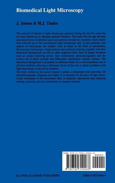
New interest in light microscopy of the last few years has not been backed up by adequate general literature. This book intends to fill the gap between specialized texts on detailed topics and general introductory booklets, mostly dealing with the use of the conventional light microscope only. In this short textbook both new developments in microscopy and basic facts of image formation will be treated, including often neglected topics such as axial resolving power, lens construction, photomicrography and correct use of phase-en interference contrast systems. Theoretical background will be dealt with as far as necessary for a well-considered application of these techniques enabling a deliberate choice for the approach of a certain problem. Over 150 illustrations (photomicrographs and diagrams) complete the information on microscopy of the nineties in the biomedical field, intended for scientists, doctors, technicians and research students. Many drawings have been contributed by the illustrator R. Kreuger; the photographic work has been executed by J. Peeterse. Secretarial assistance in preparing the manuscript was given by Ms T. M. S. Pierik. Dr M. J. Pearson has corrected the English of the final text.
Kundinnen und Kunden meinen
Verfassen Sie die erste Bewertung zu diesem Artikel
Helfen Sie anderen Kund*innen durch Ihre Meinung