Computed Tomography in Head Injuries
-
- Taschenbuch ausgewählt
- eBook
-
Sprache:Englisch
Fr. 137.00
inkl. gesetzl. MwSt.,
Beschreibung
Produktdetails
Einband
Taschenbuch
Erscheinungsdatum
21.12.2011
Verlag
Springer BerlinSeitenzahl
144
Maße (L/B/H)
24.4/17/0.9 cm
Gewicht
282 g
Auflage
Softcover reprint of the original 1st ed. 1979
Übersetzt von
F.C. Dougherty
Sprache
Englisch
ISBN
978-3-642-67423-5
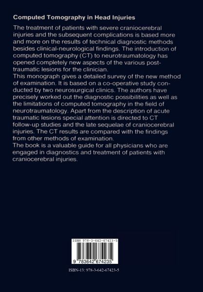
The introduction of computed tomography in the diagnosis of pathological intracranial conditions has had considerable significance in cases of cranio cerebral injury. The decisive diagnostic advantage lies in the possibility of demonstrating both gross pathological change directly as well as secondary changes in normal brain structures. Computed tomography has proved its considerable worth, especially in evaluation of patients with craniocerebral injury and its sequelae. The capabilities of CT were quickly recognized and use of the technique spread rapidly. It is likely that CT will be available within a few years in all hospitals and clinics treating patients with craniocerebral injury. We believe it appropriate to present a detailed report on our experience with CT in 1800 cases of craniocerebral injury treated in the neurosurgical departments in Miinchen-GroBhadern and Berlin-Charlottenburg over a period of five years. Both acute posttraumatic complications and late sequelae are discussed extensively. A large number of illustrations is provided in order to facilitate the reader's introduction to CT diagnosis. The great interest in our conjoint study originally published in the German language, induced us to translate this book and to update the clinical material. We wish to thank the Stiftung Volkswagenwerk, the Senat of Berlin, the Ludwig-Maximilians-Universitat in Munich and the Freie Universitat of Berlin for the generous financial support which made this study possible.
Kundinnen und Kunden meinen
Verfassen Sie die erste Bewertung zu diesem Artikel
Helfen Sie anderen Kund*innen durch Ihre Meinung