Beschreibung
Produktdetails
Einband
Taschenbuch
Erscheinungsdatum
20.03.2012
Verlag
Springer BerlinSeitenzahl
158
Maße (L/B/H)
27.9/21/1 cm
Gewicht
444 g
Auflage
Softcover reprint of the original 1st ed. 1979
Sprache
Englisch
ISBN
978-3-642-46400-3
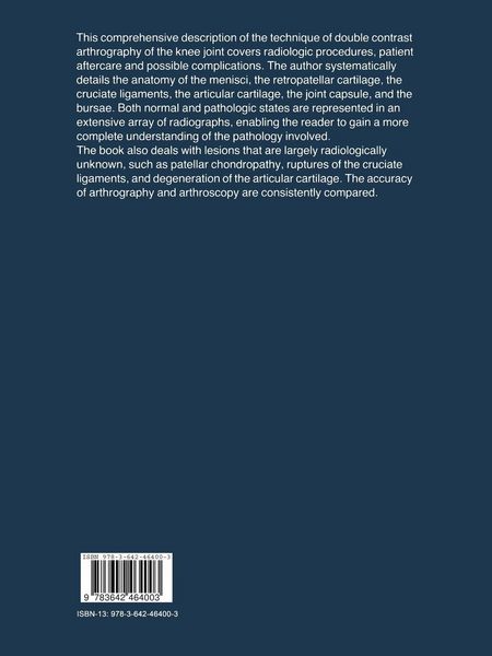
It is a great pleasure to introduce this book and its writer to the reader. Dr. Thijn has been interested in double contrast studies since he wrote his thesis on the double contrast examina tion of the colon. It would sound facetious to state that after he exhausted this field, he was in need of some other area where the same technique could be used. However, in the same exact and thorough way as in his colon studies, he has examined the knee joint. Considering that the knee is one of the most heavily taxed joints in man, with a multitude of afflictions, many of them closely connected with the age of the individual, radiological investiga tion has shown very few innovations over the decades. The true anteroposterior and lateral projections were ~ and still are ~ the mainstay of the investigation. Projections of the intercondylar fossa, and true patellar projections were used incidentally. Just prior to World War II the advent of arthrography as a double contrast investigation, as promoted by Oberholzer, was a real breakthrough.
Kundinnen und Kunden meinen
Verfassen Sie die erste Bewertung zu diesem Artikel
Helfen Sie anderen Kund*innen durch Ihre Meinung