
Nuclear Cardiology and Correlative Imaging A Teaching File
-
- Hardcover
- Taschenbuch ausgewählt
- eBook
-
Sprache:Englisch
Fr. 288.00
inkl. gesetzl. MwSt.,
Beschreibung
Produktdetails
Einband
Taschenbuch
Erscheinungsdatum
13.06.2012
Herausgeber
Joao V. Vitola + weitereVerlag
Springer UsSeitenzahl
490
Maße (L/B/H)
25.4/17.8/2.8 cm
Gewicht
960 g
Auflage
Softcover reprint of the original 1st ed. 2004
Sprache
Englisch
ISBN
978-1-4612-7392-9
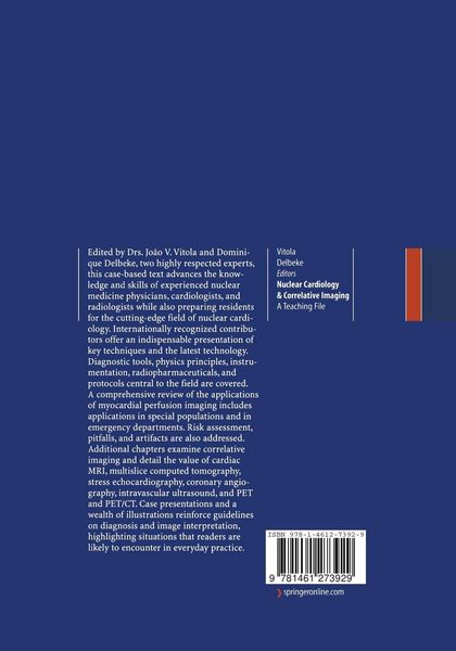
Drs. Vitola and Delbeke assembled a group of standout contributors in order to create a resource that advances the knowledge and skills of experienced nuclear cardiologists and radiologists while also preparing residents for the cutting-edge field of nuclear cardiology. Diagnostic tools, physics and instrumentation, and radiopharmaceuticals and protocols central to the field are examined. The comprehensive text covers key applications of myocardial perfusion imaging, including applications in special populations and in emergency departments. Risk assessment, pitfalls, and artefacts are addressed. Additional chapters detail the value of cardiac MRI, multislice computed tomography, stress echocardiography, and PET and PET/CT to nuclear cardiology. Practical case presentations and a wealth of illustrations reinforce instruction on diagnostic guidelines and methods.
Kundinnen und Kunden meinen
Verfassen Sie die erste Bewertung zu diesem Artikel
Helfen Sie anderen Kund*innen durch Ihre Meinung