• Produktbild: Methods of Preparation for Electron Microscopy
  • Produktbild: Methods of Preparation for Electron Microscopy

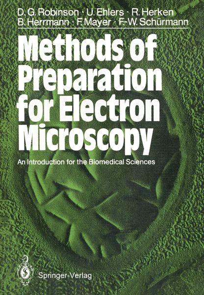
Methods of Preparation for Electron Microscopy An Introduction for the Biomedical Sciences

Fr. 137.00

inkl. gesetzl. MwSt., Versandkostenfrei


Beschreibung

Produktdetails

Einband

Taschenbuch

Erscheinungsdatum

12.06.1987

Verlag

Springer Berlin

Seitenzahl

190

Maße (L/B/H)

24.4/17/1.2 cm

Gewicht

375 g

Auflage

Softcover reprint of the original 1st ed. 1987

Übersetzt von

David G. Robinson

Sprache

Englisch

ISBN

978-3-540-17592-6

Beschreibung

Produktdetails

Einband

Taschenbuch

Erscheinungsdatum

12.06.1987

Verlag

Springer Berlin

Seitenzahl

190

Maße (L/B/H)

24.4/17/1.2 cm

Gewicht

375 g

Auflage

Softcover reprint of the original 1st ed. 1987

Übersetzt von

David G. Robinson

Sprache

Englisch

ISBN

978-3-540-17592-6

Herstelleradresse

Springer-Verlag KG
Sachsenplatz 4-6
1201 Wien
AT

Email: GPSR Kontakt

Noch keine Bewertungen vorhanden

Verfassen Sie die erste Bewertung zu diesem Artikel

Helfen Sie anderen Kundinnen und Kunden durch Ihre Meinung.

Kundinnen und Kunden meinen

Bewertungen (0)

  • Produktbild: Methods of Preparation for Electron Microscopy
  • Produktbild: Methods of Preparation for Electron Microscopy
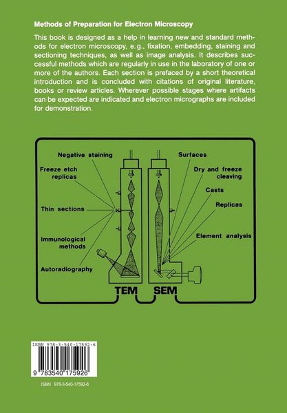
  • 1 An Introduction to Electron Microscopy (EM).- 1.1 Imaging Methods in Electron Microscopy.- 1.1.1 Conventional Transmission Electron Microscopy (TEM).- 1.1.1.1 Bright Field Electron Microscopy.- 1.1.1.2 Low Dose Transmission Electron Microscopy.- 1.1.1.3 Dark Field Electron Microscopy.- 1.1.2 Conventional Scanning Electron Microscopy (SEM).- 1.1.2.1 Imaging with Secondary and Back-Scattered Electrons.- 1.1.2.2 Scanning Electron Microscopy at Low Accelerating Voltages.- 1.2 Preparation Procedures in TEM.- 1.2.1 Overview.- 1.2.2 Structural Preservation During Fixation, Dehydration and Embedding of Biological Objects.- 1.3 Imaging Problems.- 1.3.1 On the Interpretation of TEM Images.- 1.3.2 On the Interpretation of SEM Images.- 1.4 Support Films.- 1.4.1 Grids for TEM and Their Pretreatment.- 1.4.2 Formvar Films.- 1.4.3 Collodion Films.- 1.4.4 Hydrophilisation of Films.- 1.4.5 Support Films with Holes.- 1.4.6 Carbon Films.- 2 Methods for TEM.- 2.1 Fixation, Dehydration and Embedding.- 2.1.1 Chemical Fixations.- 2.1.1.1 General Comments.- 2.1.1.2 Fixatives: Properties and Preparation.- 2.1.1.3 Composition of Fixation Solutions.- 2.1.1.4 The Fixation of Animal Cells.- 2.1.1.5 The Fixation of Plants and Microorganisms.- 2.1.1.6 The Fixation of Isolated Organelles.- 2.1.1.7 Fixing for Immunocytochemistry.- 2.1.2 Dehydration.- 2.1.3 Embedding.- 2.1.3.1 Embedding Media: General Usage and Precautions.- 2.1.3.2 Conventional Embedding.- 2.1.3.3 Water-Soluble Embedding Media.- 2.1.3.4 Embedding for Immunocytochemistry.- 2.1.3.5 Embedding Moulds and Specimen Orientation.- 2.1.3.6 Embedding of Monolayer Cell Cultures.- 2.2 Ultramicrotomy.- 2.2.1 Trimming of Blocks.- 2.2.1.1 General.- 2.2.1.2 Controlled Trimming: Production and Staining of Semi-Thin Sections.- 2.2.2 Preparing Glass Knives.- 2.2.2.1 Preparation of the Glass Strips.- 2.2.2.2 Breaking Glass Squares.- 2.2.2.3 Making Knives.- 2.2.2.4 Judging the Quality of a Glass Knife.- 2.2.2.5 Attaching Troughs.- 2.2.2.6 Storing Glass Knives.- 2.2.3 Diamond Knives and Their Care.- 2.2.4 Conventional Sectioning.- 2.2.4.1 Trough Liquids.- 2.2.4.2 Using an Ultramicrotome.- 2.2.4.3 Section Thickness.- 2.2.4.4 Picking Up Sections.- 2.2.4.5 Sectioning Problems.- 2.2.5 Cryo-ultramicrotomy.- 2.2.5.1 Freezing the Sample.- 2.2.5.2 Sectioning the Frozen Sample.- 2.2.5.3 Picking up Frozen Sections.- 2.2.6 Staining Sections.- 2.2.6.1 Staining Solutions.- 2.2.6.2 Procedure for Double Staining Sections.- 2.2.6.3 Staining Sections of Material Embedded for Immunocytochemical Purposes.- 2.2.6.4 Staining Cryosections.- 2.2.6.5 Block Staining.- 2.3 Macromolecular EM.- 2.3.1 Isolated Proteins and Protein Aggregates.- 2.3.1.1 Preparation of Specimens.- 2.3.1.2 Negative Staining Techniques.- 2.3.1.3 High Resolution Metal Shadowing.- 2.3.1.4 Preparation and Imaging of Two-Dimensional Protein Crystals.- 2.3.1.5 Making a “Tilt Series”.- 2.3.2 Isolated Nucleic Acids.- 2.3.2.1 Problems and Aims.- 2.3.2.2 Specimen Preparation.- 2.3.2.3 Spreading and Diffusion Techniques Which Employ Cytochrome c.- 2.3.2.4 “BAC” Technique.- 2.3.2.5 Partial Denaturating, Heteroduplex and R-Loop Techniques.- 2.3.3 Nucleic Acid-Protein Complexes.- 2.3.3.1 Specimen Preparation.- 2.3.3.2 Production and Staining of NA-Protein Complexes.- 2.4 Immunoelectron Microscopy (I EM).- 2.4.1 Principle Requirements.- 2.4.1.1 Antigens.- 2.4.1.2 Antibodies.- 2.4.2 Labelling of Antigens in Cells and Cell Fractions.- 2.4.2.1 Ferritin-Labelled Antibodies.- 2.4.2.2 Immunolabelling with Protein A-Gold.- 2.4.3 Localization of Protein Subunits with Specific IgG Antibodies.- 2.4.3.1 Preparation and Visualization of the Protein-Antibody Complex.- 2.5 Autoradiography.- 2.5.1 General Background.- 2.5.1.1 Physical Basis.- 2.5.1.2 Chemical Basis.- 2.5.2 Choice and Dosis of Radioactive Compounds.- 2.5.2.1 Choosing a Radioactive Precursor.- 2.5.2.2 Dosage.- 2.5.3 Working with Isotopes-Radiation Protection.- 2.5.4 Preparation of Radio-Labelled Cells/Tissues for Electron Microscopy.- 2.5.5 Photographic Emulsions and Autoradiography.- 2.5.5.1 Apparatus Required.- 2.5.5.2 Choice of Emulsion; Consequences for Resolution.- 2.5.5.3 Preparation of Sections.- 2.5.5.4 LM Autoradiography.- 2.5.5.5 Emulsion, Coating Techniques.- 2.5.6 Exposing, Developing and Fixing.- 2.5.6.1 Exposing.- 2.5.6.2 Developing and Fixing.- 2.5.6.3 Future Developments in Autoradiography.- 2.6 Freeze (Fracturing) Etching.- 2.6.1 Introduction.- 2.6.2 Freezing.- 2.6.2.1 Theoretical Background.- 2.6.2.2 Cyroprotectants.- 2.6.2.3 Supports.- 2.6.2.4 Cryogens and Freezing Methods.- 2.6.2.5 Storage of Frozen Specimens.- 2.6.3 Fracturing.- 2.6.3.1 Transfer of the Object into the Vacuum Recipient.- 2.6.3.2 The Fracturing Process.- 2.6.3.3 Fracture Planes in Biological Material.- 2.6.4 Etching.- 2.6.4.1 The Purpose of Etching.- 2.6.4.2 Theory and Practice.- 2.6.5 Shadowing and Replica Formation.- 2.6.5.1 Resistance-Heating Evaporation.- 2.6.5.2 Electron Beam Evaporation.- 2.6.5.3 Measurement of Replica Thickness.- 2.6.6 Cleaning the Replica.- 2.6.7 Artifacts in Freeze Etching.- 2.6.8 Using a Freeze-Etch Machine: a Practical Description.- 3 Methods for SEM.- 3.1 Conventional Methods of Preparation.- 3.1.1 Introduction.- 3.1.2 Specimen Size; Handling Specimens and Exposing Surfaces.- 3.1.2.1 Cleaning Surfaces.- 3.1.3 Stabilization.- 3.1.3.1 Chemical Fixations.- 3.1.3.2 Cryofixation.- 3.1.4 Dehydration.- 3.1.5 Drying.- 3.1.5.1 Critical Point Drying.- 3.1.5.2 Freeze Drying.- 3.1.6 Mounting Specimens.- 3.1.7 Increasing Conductivity.- 3.1.7.1 Sputtering.- 3.1.7.2 Evaporating.- 3.2 Storage of Specimens.- 3.3 Demonstration of Surfaces via Replicas and Casts.- 3.4 Visualization of Internal Surfaces Through Sectioning and Dry-Fracturing (Dry-Cleaving).- 3.5 Element Analysis.- 4 Evaluation of Micrographs.- 4.1 Morphometry.- 4.1.1 Problems and Solutions.- 4.1.2 Measurement: Some General Points.- 4.1.3 Stereology: General Principles.- 4.1.4 Collection and Evaluation of Data; Statistical Treatments.- 4.2 Averaging and Image Reconstruction.- 4.2.1 General.- 4.2.2 Markham Rotation.- 4.2.3 Principles of Light Optical Diffraction.- 4.2.4 Principles of Computer-Assisted Image Reconstruction.- Appendix: Buffers in Electron Microscopy.