Atlas of Laser Scanning Ophthalmoscopy
-
- Hardcover ausgewählt
- Taschenbuch
-
Sprache:Englisch
Fr. 138.00
inkl. gesetzl. MwSt.,
Beschreibung
Produktdetails
Einband
Gebundene Ausgabe
Erscheinungsdatum
03.12.2003
Abbildungen
w. 100 col. Illustrationen
Verlag
Springer BerlinSeitenzahl
170
Maße (L/B/H)
26/19.3/1.1 cm
Gewicht
597 g
Auflage
1. Auflage
Sprache
Englisch
ISBN
978-3-540-01868-1
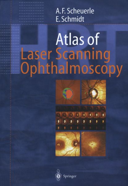
Glaucoma remains one of the leading causes of blindness. Laser scanning tomography has gained an indispensable role in the ophthalmologic diagnosis, especially in the long-term follow-up of glaucoma. Confocal laser scanning ophthalmoscopy provides key insights into the three-dimensional anatomy of the optic disc in vivo. This unique atlas contains superb images of all clinically relevant diseases diagnosed by current models of the Heidelberg Retina Tomograph. It correlates classical diagnostic tools like perimetry and fundus photography with state-of-the-art studies including digital retinal angiography, optical coherence tomography and laser scanning tomography. Special features include the illustrated coverage of:
Diseases of the optic nerve head; different types and stages of glaucoma, non-glaucomatous neuropathy and papilledema; automated classification procedures for the detection of glaucoma; strategies for the interpretation of follow-up results in optic disc monitoring.
Macular diseases: the shortly released Retina Module expands the diagnostic spectrum of laser scanning ophthalmoscopy significantly, adding measuring and monitoring of diabetic and cystoid macular edema.
This atlas is the most comprehensive up-to-date reference of laser scanning ophthalmoscopy available, ideal for residents and general ophthalmologists who want to enhance their diagnostic skills.
Noch keine Bewertungen vorhanden
Verfassen Sie die erste Bewertung zu diesem Artikel
Helfen Sie anderen Kundinnen und Kunden durch Ihre Meinung.