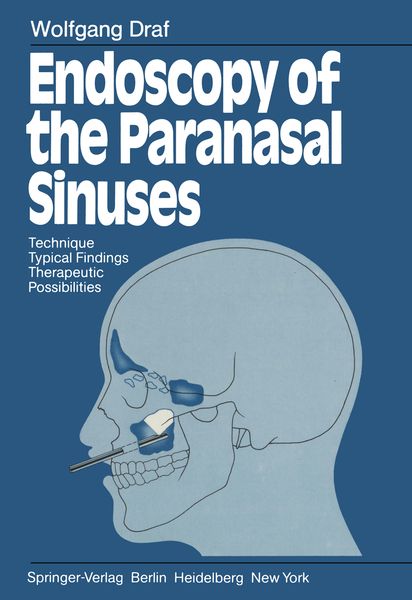
Endoscopy of the Paranasal Sinuses Technique · Typical Findings Therapeutic Possibilities
-
- Englisch ausgewählt
Fr. 137.00
inkl. gesetzl. MwSt.,
Beschreibung
Produktdetails
Einband
Taschenbuch
Erscheinungsdatum
18.11.2011
Abbildungen
X, 102 p.
Verlag
Springer BerlinSeitenzahl
102
Maße (L/B/H)
24.4/17/0.7 cm
Gewicht
216 g
Übersetzt von
William E. Pohl
Sprache
Englisch
ISBN
978-3-642-68414-2
The exponential explosion of diagnostic techniques that is the hallmark of Twentieth Century medicine often makes us forget that the human perceptual senses are still the simplest, most reliable, and most readily available means of recognizing disease and detecting abnormalities in the human body. Improvements in optical telescopes and in glass rod light transmission have enables us to look directly into the human body, and they are indispensable to modern endoscopy. The present monograph is a product of many years of diligent search and tireless work by Professor WOLFGANG DRAF in the field of endoscopy of the parana sal sinuses. Scholastic curiosity, pursuit of academic excellence, and a fervent desire to give patients prompt and lasting relief of symptoms caused by paranasal sinus disease were the author's initial motivations. As time went on and the methodology became more refined, he ventured from the field of endoscopic diagnostic and photographic documentation into the area of endoscopic therapy for selective diseases of maxillary, frontal, and sphenoid sinuses. Many of the instruments and most of the optical and photo graphic equipment and accessories have been developed and improved in parallel with the author's professional career; indeed, many of these technical innovations were a product of Professor DRAF'S keen and methodical research.
Noch keine Bewertungen vorhanden
Verfassen Sie die erste Bewertung zu diesem Artikel
Helfen Sie anderen Kundinnen und Kunden durch Ihre Meinung.