KI-Beratung Beta

Produktbild: Comprehensive Atlas of High-Resolution Endoscopy and Narrowband Imaging

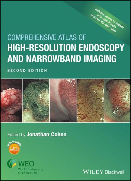
Comprehensive Atlas of High-Resolution Endoscopy and Narrowband Imaging

Aus der Reihe Contesting the Past

Fr. 269.00

inkl. gesetzl. MwSt., Versandkostenfrei


Beschreibung

Previous edition won First Prize in the Gastroenterology category of the 2008 BMA Medical Book Competition   High-resolution endoscopy and narrowband imaging have revolutionized the field. Edited by a gastroenterologist with a reputation for delivering outstanding material, this new edition of an award-winning...

Beschreibung

Previous edition won First Prize in the Gastroenterology category of the 2008 BMA Medical Book Competition
 
High-resolution endoscopy and narrowband imaging have revolutionized the field. Edited by a gastroenterologist with a reputation for delivering outstanding material, this new edition of an award-winning atlas provides you with an outstanding collection of images, videos, and expert diagnostic guidance to enhance your decision making.
 
To accelerate your learning, Dr. Cohen offers more than 2000 endoscopic images, emphasizing conditions for which NBI is particularly useful - such as finding dysplasia in Barrett's mucosa, and diagnosing adenomatous colon polyps - and providing exceptional preparation for the future of endoscopy practice, with a broad new look at normal and abnormal findings throughout the GI tract.
 
The book is divided into three main parts:
* The basics of NBI
* Clinical applications of NBI
* Atlas of 1600 color images, broken into sections on the pharynx and esophagus, stomach, small intestine, and colon, including correlating histopathology and multiple examples of key pathologies
 
The accompanying website features more than 85 video clips containing over 3 hours of annotated video, to give you a complete sense of how HRE and NBI work and look in real time, including during therapeutic procedures.
 
All of the over 1000 new images appear in much brighter color, reflecting the advance in scope technology since the first edition. New chapters have been added to present the data supporting increased use of NBI in optical diagnosis and in the context of therapeutic procedures. For the first time, brilliant images of the bile duct and pancreas are included as the imaging revolution has expanded to reach these new locations. This spectacular new imaging modality promises to enhance endoscopic decision making in real time, facilitate therapeutic maneuvers, make tissue sampling more precise, and make resection of mucosal neoplasia more complete.
 
Expertly guiding you through the latest advances, this book facilitates your mastery of the field, and provides an up-to-date reference for gastroenterologists and endoscopists to improve their practice.

Produktdetails

Einband

Gebundene Ausgabe

Erscheinungsdatum

24.04.2017

Herausgeber Jonathan Cohen
Verlag John Wiley & Sons Inc

Produktdetails

Einband

Gebundene Ausgabe

Erscheinungsdatum

24.04.2017

Herausgeber Jonathan Cohen
Verlag John Wiley & Sons Inc
Seitenzahl

576

Maße (L/B/H)

25.1/18.9/3.2 cm

Gewicht

1552 g

Auflage

2nd edition

Sprache

Englisch

ISBN

978-1-118-70593-3

Herstelleradresse

Libri GmbH
Europaallee 1
36244 Bad Hersfeld
DE

Email: gpsr@libri.de

Noch keine Bewertungen vorhanden

Verfassen Sie die erste Bewertung zu diesem Artikel

Helfen Sie anderen Kundinnen und Kunden durch Ihre Meinung.

Kundinnen und Kunden meinen

Bewertungen (0)

Die Leseprobe wird geladen.
  • Produktbild: Comprehensive Atlas of High-Resolution Endoscopy and Narrowband Imaging
  • Preface vi
     
    Acknowledgments viii
     
    Contributors ix
     
    About the Companion Website xii
     
    List of videos illustrating key procedures xiii
     
    Part 1: The Basics of NBI
     
    1 Narrow band imaging: historical background and basics for its development 3
    Shigeaki Yoshida
     
    2 An introduction to high-resolution endoscopy and narrow band imaging 7
    Kazuhiro Gono
     
    3 Getting started with narrow band imaging 16
    Neil Gupta, Ajay Bansal, and Prateek Sharma
     
    Part 2: Application of High-Resolution Endoscopy and NBI in Diagnosis and Therapy
     
    Section 1: Pharynx and Esophagus
     
    4 Detection and superficial cancer in oropharyngeal and hypopharyngeal mucosal sites and the value of NBI in qualitative diagnosis 27
    Manabu Muto, Atsushi Ochiai, and Shigeaki Yoshida
     
    5 Magnifying endoscopic diagnosis of tissue atypia and cancer in vasion depth in the area of pharyngoesophageal squamous epithelium by NBI enhanced magnification image: IPCL pattern classification 39
    Robert Bechara and Haruhiro Inoue
     
    6 Applications of NBI HRE abd preliminary data: Barrett's esophagus and adenocarcinoma 60
    Anne-Fré Swager, Prateek Sharma, Jacques J. Bergman and Wouter L. Curvers
     
    Section 2: Stomach and Duodenum
     
    7 Clinical application of magnification endoscopy with NBI in the stomach and the duodenum 75
    Kenshi Yao, Takashi Nagahama, Fumihito Hirai, Suketo Sou, Toshiyuki Matsui, Hiroshi Tanabe, Akinori Iwashita, Philip Kaye and Krish Ragunath
     
    8 Magnifying endoscopy with NBI in the diagnosis of superficial gastric neoplasia and its application for endoscopic submucosal dissection 94
    Mitsuru Kaise
     
    Section 3: Colon
     
    9 Optical chromoendoscopy using NBI during screening colonoscopy: its usefulness and application 109
    Yasushi Sano and Shigeaki Yoshida
     
    10 Keys to effective performance of resect and discard NBI 131
    Douglas K. Rex
     
    11 The significance of NBI observation for inflammatory bowel diseases 146
    Takayuki Matsumoto, Tetsuji Kudo, Mitsuo Iida, and James East
     
    Section 4: New Horizons for High-Resolution Endoscopy and Narrow band Imaging
     
    12 NBI and high-resolution endoscopy in the bile duct and pancreas 163
    Takao Itoi
     
    13 Applications in the therapeutic endoscopy: impact on timing and completeness of mucosal ablation and resections 174
    Michael J. Bartel, Lijia Jiang, and Michael B. Wallace
     
    Part 3: Atlas of Images and Histopathologic Correlates
     
    14 Pharynx and esophagus atlas 185
     
    15 Stomach atlas 257
     
    16 Small intestine atlas 317
     
    17 Colon atlas 357
     
    Index 553