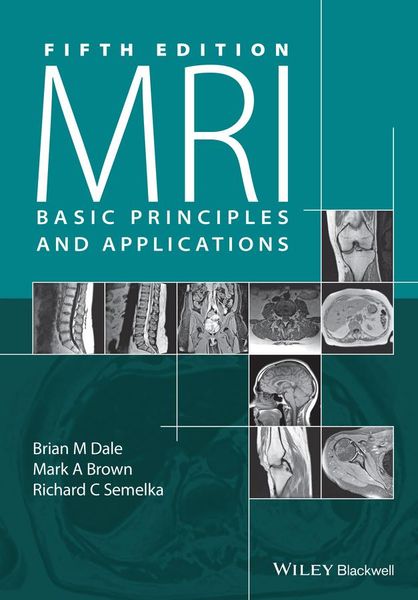
Produktbild: MRI

MRI Basic Principles and Applications

Fr. 99.90

inkl. gesetzl. MwSt., Versandkostenfrei


Beschreibung

Produktdetails

Einband

Taschenbuch

Erscheinungsdatum

19.10.2015

Verlag

John Wiley & Sons Inc

Seitenzahl

248

Maße (L/B/H)

24.8/17.2/1.4 cm

Gewicht

481 g

Auflage

5th Revised edition

Sprache

Englisch

ISBN

978-1-119-01305-1

Beschreibung

Produktdetails

Einband

Taschenbuch

Erscheinungsdatum

19.10.2015

Verlag

John Wiley & Sons Inc

Seitenzahl

248

Maße (L/B/H)

24.8/17.2/1.4 cm

Gewicht

481 g

Auflage

5th Revised edition

Sprache

Englisch

ISBN

978-1-119-01305-1

Herstelleradresse

Libri GmbH
Europaallee 1
36244 Bad Hersfeld
DE

Email: GPSR Kontakt

Noch keine Bewertungen vorhanden

Verfassen Sie die erste Bewertung zu diesem Artikel

Helfen Sie anderen Kundinnen und Kunden durch Ihre Meinung.

Kundinnen und Kunden meinen

Bewertungen (0)

Die Leseprobe wird geladen.
  • Produktbild: MRI
  • Preface, ix

    ABR study guide topics, xi

    1 Production of net magnetization 1

    1.1 Magnetic fields 1

    1.2 Nuclear spin 2

    1.3 Nuclear magnetic moments 4

    1.4 Larmor precession 4

    1.5 Net magnetization 6

    1.6 Susceptibility and magnetic materials 8

    2 Concepts of magnetic resonance 10

    2.1 Radiofrequency excitation 10

    2.2 Radiofrequency signal detection 12

    2.3 Chemical shift 14

    3 Relaxation 17

    3.1 T1 relaxation and saturation 17

    3.2 T2 relaxation, T2* relaxation, and spin echoes 21

    4 Principles of magnetic resonance imaging - 1 26

    4.1 Gradient fields 26

    4.2 Slice selection 28

    4.3 Readout or frequency encoding 30

    4.4 Phase encoding 33

    4.5 Sequence looping 35

    5 Principles of magnetic resonance imaging - 2 39

    5.1 Frequency selective excitation 39

    5.2 Composite pulses 44

    5.3 Raw data and image data matrices 46

    5.4 Signal-to-noise ratio and tradeoffs 47

    5.5 Raw data and k-space 48

    5.6 Reduced k-space techniques 51

    5.7 Reordered k-space filling techniques 54

    5.8 Other k-space filling techniques 56

    5.9 Phased-array coils 58

    5.10 Parallel acquisition methods 60

    6 Pulse sequences 65

    6.1 Spin echo sequences 67

    6.2 Gradient echo sequences 70

    6.3 Echo planar imaging sequences 75

    6.4 Magnetization-prepared sequences 77

    7 Measurement parameters and image contrast 86

    7.1 Intrinsic parameters 87

    7.2 Extrinsic parameters 89

    7.3 Parameter tradeoffs 91

    8 Signal suppression techniques 94

    8.1 Spatial presaturation 94

    8.2 Magnetization transfer suppression 96

    8.3 Frequency-selective saturation 99

    8.4 Nonsaturation methods 101

    9 Artifacts 103

    9.1 Motion artifacts 103

    9.2 Sequence/Protocol-related artifacts 105

    9.3 External artifacts 119

    10 Motion artifact reduction techniques 126

    10.1 Acquisition parameter modification 126

    10.2 Triggering/Gating 127

    10.3 Flow compensation 132

    10.4 Radial-based motion compensation 134

    11 Magnetic resonance angiography 135

    11.1 Time-of-flight MRA 137

    11.2 Phase contrast MRA 141

    11.3 Maximum intensity projection 144

    12 Advanced imaging applications 147

    12.1 Diffusion 147

    12.2 Perfusion 153

    12.3 Functional brain imaging 156

    12.4 Ultra-high field imaging 158

    12.5 Noble gas imaging 159

    13 Magnetic resonance spectroscopy 162

    13.1 Additional concepts 162

    13.2 Localization techniques 167

    13.3 Spectral analysis and postprocessing 169

    13.4 Ultra-high field spectroscopy 173

    14 Instrumentation 177

    14.1 Computer systems 177

    14.2 Magnet system 180

    14.3 Gradient system 182

    14.4 Radiofrequency system 184

    14.5 Data acquisition system 186

    14.6 Summary of system components 187

    15 Contrast agents 189

    15.1 Intravenous agents 190

    15.2 Oral agents 195

    16 Safety 196

    16.1 Base magnetic field 197

    16.2 Cryogens 197

    16.3 Gradients 198

    16.4 RF power deposition 198

    16.5 Contrast media 199

    17 Clinical applications 200

    17.1 General principles of clinical MR imaging 200

    17.2 Examination design considerations 202

    17.3 Protocol considerations for anatomical regions 203

    17.4 Recommendations for specific sequences and clinical situations 218

    References and suggested readings 222

    Index 225