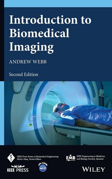
Introduction to Biomedical Imaging
Aus der Reihe
IEEE Press Series on Biomedical Engineering
-
Sprache:Englisch
-
Verlag:John Wiley & Sons Inc
Fr. 193.00
inkl. gesetzl. MwSt.,
Beschreibung
Produktdetails
Einband
Gebundene Ausgabe
Erscheinungsdatum
04.10.2022
Verlag
John Wiley & Sons IncSeitenzahl
384
Maße (L/B/H)
2.2/15.2/22.9 cm
Gewicht
615 g
Auflage
2. Auflage
Sprache
Englisch
ISBN
978-1-119-86771-5
Introduction to BiomedicalImaging
A state-of-the-art exploration of the foundations and latest developments in biomedical imaging technology
In the newly revised second edition of Introduction to Biomedical Imaging, distinguished researcher Dr. Andrew Webb delivers a comprehensive description of the fundamentals and applications of the most important current medical imaging techniques: X-ray and computed tomography, nuclear medicine, ultrasound, magnetic resonance imaging, and various optical-based methods. Each chapter explains the physical principles, instrument design, data acquisition, image reconstruction, and clinical applications of its respective modality.
This latest edition incorporates descriptions of recent developments in photon counting CT, total body PET, superresolution-based ultrasound, phased-array MRI technology, optical coherence tomography, and iterative and model-based image reconstruction techniques. The final chapter discusses the increasing role of artificial intelligence/deep learning in biomedical imaging. The text also includes a thorough introduction to general image characteristics, including discussions of signal-to-noise and contrast-to-noise.
Perfect for graduate and senior undergraduate students of biomedical engineering, Introduction to Biomedical Imaging, 2nd Edition will also earn a place in the libraries of medical imaging professionals with an interest in medical imaging techniques.
A state-of-the-art exploration of the foundations and latest developments in biomedical imaging technology
In the newly revised second edition of Introduction to Biomedical Imaging, distinguished researcher Dr. Andrew Webb delivers a comprehensive description of the fundamentals and applications of the most important current medical imaging techniques: X-ray and computed tomography, nuclear medicine, ultrasound, magnetic resonance imaging, and various optical-based methods. Each chapter explains the physical principles, instrument design, data acquisition, image reconstruction, and clinical applications of its respective modality.
This latest edition incorporates descriptions of recent developments in photon counting CT, total body PET, superresolution-based ultrasound, phased-array MRI technology, optical coherence tomography, and iterative and model-based image reconstruction techniques. The final chapter discusses the increasing role of artificial intelligence/deep learning in biomedical imaging. The text also includes a thorough introduction to general image characteristics, including discussions of signal-to-noise and contrast-to-noise.
Perfect for graduate and senior undergraduate students of biomedical engineering, Introduction to Biomedical Imaging, 2nd Edition will also earn a place in the libraries of medical imaging professionals with an interest in medical imaging techniques.
Noch keine Bewertungen vorhanden
Verfassen Sie die erste Bewertung zu diesem Artikel
Helfen Sie anderen Kundinnen und Kunden durch Ihre Meinung.